Redefining the Vision of Skin Care

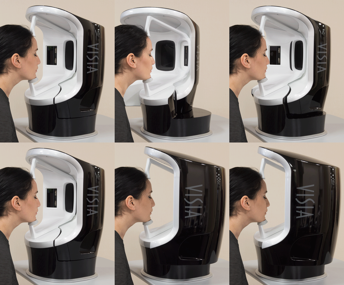
The 7th Generation VISIA® Skin Analysis System delivers a significantly improved experience for aesthetic and skin care consultations. A newly designed capture module rotates smoothly around the subject, greatly simplifying the imaging process while providing greater comfort for the client. Updated software allows faster image capture with automatic skin type classification, refined facial feature detection and more.

VISIA® Skin Analysis

Easily Capture High Quality, Standardized Facial Images

VISIA's multi-point positioning system and live image overlay make it easy to capture perfectly registered images to document progress over time. Rotating capture module easily captures left, right and frontal facial views.

VISIA Skin Analysis System capturing a clients face from the frontal view
IntelliFlash®, cross-polarized and UV lighting are used to record and measure surface and subsurface skin conditions. UV photography provides the most complete data set available for sun damage assessment and analysis, including UV fluorescence imaging to reveal porphyrins.
Canfield's RBX® Technology separates the unique color signatures of Red and Brown skin components for unequaled visualization of conditions that result in color concentration, such as spider veins, hyperpigmentation, inflammation and other conditions.

Spots

Typically brown or red skin marks, distinguishable by the distinct color and contrast from the background skin tone. Spots vary in size and shape, and are normally visible to the naked eye.

Spots

Wrinkles

Furrows, folds or creases in the skin that can increase as a result of sun exposure and are associated with a decrease in skin elasticity.

Wrinkles

Texture

Raised and depressed areas that indicate variations on the skin surface affecting skin smoothness.

Textures

Pores

Circular surface openings of sweat gland ducts. Due to shadowing, enlarged pores appear darker than the surrounding skin tone and are identified by the darker color and circular shape.

Pores

UV Spots

The absorption of UV light by epidermal melanin just below the skin surface results in the display and detection of UV spots that can result from sun damage.

UV Spots

Brown Spots

Pigmentation and discoloration on and beneath the surface of the skin.

Brown Spots

Red Areas

Concentrated darker red areas, which may be related to a variety of conditions such as inflammation or spider veins.

Red Areas

Porphyrins

Bacterial excretions that can become lodged in pores.

Porphyrins

Clear Communication

Select personalized recommendations from VISIA's built-in library of skin care products and treatments. Your staff will sell rejuvenation and skin care treatments more effectively with clear recommendations combined with the visual impact of VISIA Skin Analysis. Your recommendations also appear in the printed report and in the ViewMyConsult web portal.

With ViewMyConsult, stay connected with clients as they consider the possibilities of aesthetic procedures. Clients can view their pictures, treatment recommendations, and treatment progress at home using a secure, password-protected web portal.

Personalized Printed Reports record easy to understand information for use in client education and treatment planning. Reports also serve as a valuable aid for increasing referrals.

Quantitative Analysis & Visual Assessment


A.I. Wrinkle Algorithm

A.I. Wrinkle Algorithm

New in 8.4, the first ever use of A.I. in the VISIA Experience. The new wrinkle algorithm significantly improves detection and accuracy of fine lines and wrinkles.

UV Sepia Tone New with VISIA 8.3.2

UV Sepia Tone New with VISIA 8.3.2

New with 8.3.2, the optional sepia tone for displaying UV images is an alternative option for aesthetically pleasing UV images.


Positioning Grid Lines New with VISIA 8.3.2

Positioning Grid Lines New with VISIA 8.3.2

New with 8.3.2, the optional grid preview assists with patient positioning and photo repeatability.

Injectables Simulation

Injectables Simulation

New with VISIA 8.2, the injectables simulation may be applied to the degree desired for each region to simulate treatments.


iPad App Update

iPad App Update

All of VISIA's analytic and communication tools are available for the iPad, enabling VISIA Consultations from anywhere in the office. Now introducing TruSkin Age® and Compare!

Advanced Aging Simulation

Advanced Aging Simulation

New with VISIA'S 8.1 Software. Simulate how clients might appear from their current age to 80 years old (Front facing images only)


TruSkin Age®

TruSkin Age®

Determines the patient's overall skin condition and age, providing a revolutionary new metric to guide the selection of skin care products and treatment options.

Updated RBX® Technology

Updated RBX® Technology

NEW with VISIA 8.1 Software. Updated RBX® technology provides a more refined visualization of sub-surface melanin and vascular conditions for all skin types and ethnicities.


Percentile Scores

Percentile Scores

VISIA's patented comparison to norms analysis uses the world's largest skin feature database to grade your patient's skin relative to others of the same age and skin type. Measure spots, wrinkles, texture, pores, UV spots, brown spots, red areas, and porphyrins.

Meaningful Comparisons

Meaningful Comparisons

Compare results side by side for any combination of views, features or time points, including graphs and numerical data. Zoom and pan images in tandem for clear and easy comparisons.


Eyelash Analysis

Eyelash Analysis

Evaluates the results of lash improvement treatments with numerical assessments and graphic visualizations.

Loupe Tool

Loupe Tool

Target a selected area of the image for maximum impact. Zoom within, expand or shrink the loupe using a mouse or touchscreen.


3D Viewer

3D Viewer

Visualize the skin's surface in three dimensions from any angle, rendered in natural skin tone, color relief, or gray scale.

Aging Simulation

Aging Simulation

Simulate the aging process to show clients older or younger by 5-7 years for spots, wrinkles or both.


Capture-Only Mode

Capture-Only Mode

Zip through repeatable facial imaging for photo documentation without pausing to process the images. Analysis may be performed later, if desired.

Skin Type Detection

Skin Type Detection

Automatic detection of the client’s skin type for streamlined image processing and feature analysis.


iPad Consultation App

iPad Consultation App

All of VISIA's analytic and communication tools are available for the iPad, enabling VISIA Consultations from anywhere in the office.

ViewMyConsult® Integration

ViewMyConsult® Integration

Stay connected as your clients consider the possibilities of aesthetic treatments. By accessing a secure, password-protected web portal, clients can view their photographs, treatment recommendations, and progress of treatments in the privacy of their own home.


Product Library

Product Library

The cloud-based Product Recommendations Library enables you to easily add your products, treatments and services to your client records from the facial imager software. The centralized product library resource is managed directly by the manufacturers.

Mirror® Integration

Mirror® Integration

A direct interface with Canfield's Mirror medical imaging software (available separately), which communicates the benefits of resurfacing and injectables.


Canfield Care® Available

Canfield Care® Available

Unlimited technical support, hardware warranty, training webinars, software upgrades and ViewMyConsult access.

Tech Specs

Specifications

VISIA Facial Imaging Booth
  • Automatic focus
  • Automated white balance correction
  • Capture module positions: Left 33°, Center 0°, Right 33°
  • Headrest: Adjustable vertically three positions
  • Chinrest Height: 8.8 in (22.3 cm) from table
  • Headrest Height: 16 in (40.6 cm) from table
  • Multi-spectral Imaging
    • Standard light IntelliFlash
    • Cross Polarized flash
    • Ultraviolet lighting
VISIA Facial Imaging Booth - Size & Weight
  • Centered: 13 x 14.2 x 20.9 in (32.9 x 36 x 53.1 cm)
  • Total footprint with 3 position swing: 15 x 22.9 x 20.9 in (38.1 x 58.2 x 53.1 cm)
  • Weight: 31 lbs. (14 kg)
Power
  • Voltage: 100 to 240 Volts AC (+/- 10%)
  • Frequency: 50/60 Hz (+/- 3 Hz)
  • Current: 3 A (rms)

Requirements

Workstation
  • 16 GB RAM
  • Intel i5 or AMD equivalent processor required, 2.2 GHz or faster. ARM-based PC processors are not supported
  • Minimum 10 GB available disk space for installing application
  • 250 GB available disk space recommended for data
  • Monitor: 1280 x 900 (4K monitors supported at maximum 150% magnification)
  • USB Ports: 2 dedicated ports (USB 2.0)
  • 1 Gbps wired LAN connectivity (Wi-Fi not supported)
  • Windows 10 or 11 (Professional 64 Bit) - Recommended
  • Windows Home Edition & Windows VISTA – NOT SUPPORTED
VISIA iPad App
  • Minimum support of 802.11n or later protocol by the intra-office Wi-Fi router
  • Requires iPad capable of running the current version of iOS
SQL Server
  • Windows Server 2019 64-bit and 2022 64-bit
  • Microsoft SQL Server 2017 and 2019 (Express edition is installed by default)
  • 2 GHz or faster processor, Intel i5
  • 16 GB RAM (or higher)
  • 1 Gbps wired LAN connectivity (Wi-Fi not supported)
  • 300 GB HD, 7200 rpm minimum
  • RAID 1 - NOT SUPPORTED
  • Space required dependent upon patient volume and camera used/settings
  • Regularly scheduled backups are highly recommended
Notes
  • Users must be logged on with local administrator privileges.
  • Serial Numbers below 947249 are no longer supported. Contact Canfield for trade in / upgrade options.
  • The serial number is located next to the USB port on the rear panel.
  • We do not support using a server class OS as a workstation.
  • WAN Connectivity is NOT supported.

Frequently Asked Questions

What are the definitions of the various skin features and how are the features detected?

Spots : Spots are typically brown or red skin lesions including freckles, acne scars, hyper-pigmentation and vascular lesions. Spots are distinguishable by their distinct color and contrast from the background skin tone. Spots vary in size and generally have a circular shape.

Pores : Pores are the circular surface openings of sweat gland ducts. Due to shadowing, pores appear darker than the surrounding skin tone and are identified by their darker color and circular shape. The VISIA system distinguishes pores from spots based on size; by definition, the area of a pore is much smaller than a spot.

Wrinkles : Wrinkles are furrows, folds or creases in the skin, which increase in occurrence as a result of sun exposure, and are associated with decreasing skin elasticity. This skin feature has the greatest variability from image to image as it is highly dependent upon the facial expression of the client. Wrinkles are identified by their characteristic long, narrow shape.

Texture :Texture is primarily an analysis of skin smoothness. Texture measures skin color and smoothness by identifying gradations in color from the surrounding skin tone, as well as peaks (shown in yellow) and valleys (shown in blue) on the skin surface that indicate variations in the surface texture.

Porphyrins : Porphyrins are bacterial excretions that can become lodged in pores and lead to acne. Porphyrins fluoresce in UV light and exhibit circular white spot characteristics.

UV Spots : UV spots occur when melanin coagulates below the skin surface as a result of sun damage. UV spots are generally invisible under normal lighting conditions. The selective absorption of the UV light by the epidermal melanin enhances its display and detection by VISIA.

Red Areas : Red Areas represent a potential variety of conditions, such as acne, inflammation, Rosacea or spider veins. Blood vessels and hemoglobin contained in the papillary dermis, a sub-layer of skin, give these structures their red color, which is detected by the RBX Technology in VISIA. Acne spots and inflammation vary in size but are generally round in shape. Rosacea is usually larger and diffuse compared to acne, and spider veins usually are short, thin and can be interconnected in a dense network.

Brown spots\ : Brown Spots are lesions on the skin such as hyper-pigmentation, freckles, lentigines, and melasma. Brown Spots occur from an excess of Melanin. Melanin is produced by melanocytes in the bottom layer of the epidermis. Brown Spots produce an uneven appearance to the skin, and are detected in VISIA by RBX.

What factors influence the results of the image session?

A number of factors contribute to the actual scores generated by the VISIA complexion analysis algorithms, including the condition of the skin (cleaned vs. fresh make-up, old make-up, time-of-day), head registration within the booth, facial expression (particularly for wrinkles), stray hair, glare areas resulting from oily skin, etc. Note that the percentile scores were generated for subjects with cleaned skin prior to their imaging session, so results may vary if make-up is NOT removed prior to a patient's VISIA session.

What is the definition for each of the three types of skin conditions? How does VISIA use this information?

"Clean" is defined as skin that has been recently cleansed and without make-up or skin conditioners applied, "Fresh Make-up" is defined as make-up recently applied (roughly within the past hour), "Other" covers all other cases. No change is made in terms of either taking the photo or in the analysis process based upon the skin condition selected.

This information is recorded in the patient's record for each session and allows scores to be properly interpreted if images were captured under different conditions session-to-session (i.e., one time image taken with clean skin, the next time with make-up). Also, images that are "clean" can be considered as valid input to further build the database.

Which data display option is best for our practice to use?

Three data display options are available depending upon the needs and preferences of the practice, and can either be selected from within VISIA's Session Options tab or by right clicking on any data display bar or graph.

Feature Counts : Feature Counts provide a count of the number of discrete instances of the feature being evaluated, without regard to the size or intensity of each instance. Feature Counts can be used to track treatment progress when a reduction in the number of discrete instances of a particular feature is desired.

Absolute Scores : Absolute Scores provide a comprehensive measurement of the impact that the feature has on the client's complexion. Absolute Scores factor in the total size and area as well as intensity of detected instances of the feature being analyzed. Absolute Scores can be used to track treatment progress when the size and intensity of instances of a feature are the most relevant indicator of treatment effectiveness.

Percentiles : Percentiles provide a context in which to evaluate a client's complexion analysis results by presenting a comparison of the individual's Absolute Scores to those of people with similar characteristics. Percentiles are useful in providing a baseline assessment of the overall condition of the client's complexion.

What clinical methods were used to validate VISIA?

The Procter & Gamble complexion analysis software used in VISIA was developed in the late 1990s and has undergone extensive lab and field-testing. Since 1998, P&G has deployed their complexion analysis software as a sales tool to promote their Olay and SKII brands of cosmetic products. As such, the P&G software was never intended for clinical trials. However, the VISIA complexion analysis algorithms themselves are supported by an extensive set of research papers describing the analysis techniques and results across various skin types.

What is the wavelength used for the UV photography?

The wavelength used by the UV flashes is 365 nm (peak).

Should the patient's eyes be open or closed when the picture is taken?

It is strongly recommended that the eyes be closed for the UV photos given the intensity of the UV flashes. For standard photos, the eyes can be either open or closed, although more aesthetically pleasing images are achieved with the eyes open. Because the mask is created using the standard photo (in which the patient's eyes are typically open), there is no need to be concerned that eyelashes will be included inadvertently in UV analysis, even though the patient's eyes are closed.

Why isn't the porphyrin count reduced after cleansing?

There are several potential reasons why the porphyrin count for a particular patient may not be reduced-or worse case, increase, after cleansing his/her skin. First, certain types of paper/tissue that might have been used to dry the face after cleansing could have a high content of lint particles. Lint will actually fluoresce under UV light. Subject matter expert Greg Hillebrand, Ph.D., offers the further explanation that "in some situations, the true porphyrin fluorescence of an initial image could be quenched by something that the subject had on his/her unclean face, and then after removal of this interfering quenching agent, a higher level of fluorescence could be observed. Finally, the simple cleansing of the skin with a normal everyday facial cleanser is probably not going to change the true number of pores that show porphyrin fluorescence. It has been reported that washing with a cleanser containing benzoyl peroxide will, over time, reduce the number of pores. Also, using strips or other devices to physically remove the contents of the pores will help to reduce the fluorescence as well."

Is there correlation between porphyrins and sun damage?

According to subject matter expert Greg Hillebrand, Ph.D., there may be a real correlation between the amount of porphyrins and skin aging as described in the following technical excerpt that offers the best understanding we have today on the matter: "In pilosebaceous follicles, porphyrins are produced by propionibacterium acnes (P. acnes) that are normal inhabitants of the human skin and the pilosebaceous ducts. Porphyrins are highly fluorescent pores containing P. acnes and the associated porphyrins appear red-orange when facial skin is illuminated with long wavelength UVA radiation. The intensity of the follicular fluorescence and its extent of facial involvement is known to be proportional to the density of P. acnes. Porphyrin fluorescence appears first on the nose and chin, and then increases in incidence through adulthood and declines after the age of 50, possibly reflecting the rate of sebum secretion. Porphyrins may also have cytotoxic and comedogenic potential. With exposure to the sun, porphyrins can release singlet oxygen that in turn can oxidize lipids (lipid peroxidation) in the skin yielding irritating and cytotoxic lipid peroxides that can cause skin damage. Thus, the presence of porphyrins in the follicular pores may enhance or accelerate hyperpigmentation, wrinkling and other signs of photo-aging by sun exposure."

Support

Canfield Support
  • 1-year limited warranty, including support & upgrades
  • Includes advanced replacement, round trip shipping in US
  • Includes software feature improvements and enhancements
  • Canfield Care available

Warranty

VISIA Facial Imaging Booth & Software
  • 1-year limited warranty, including support & upgrades
  • Includes advanced replacement, round trip shipping in US
  • Includes software feature improvements and enhancements
  • Canfield Care available
Dell All-in-One Computer (VISIA Deluxe)
  • 3-year limited warranty, including on-site, next day service from Dell, in the US

Training

VISIA Training
  • Live, web-based training, two-hour session included
  • Additional on-site training by Canfield is available, contact us for pricing and additional information.
  • Unlimited Webinars are included at no charge during the first year or with Canfield Care ($250 value per webinar)